Undescended testicle is the condition where the testicles are not in the scrotum where they should be. It is noticed during routine physical examinations of newborn male babies.
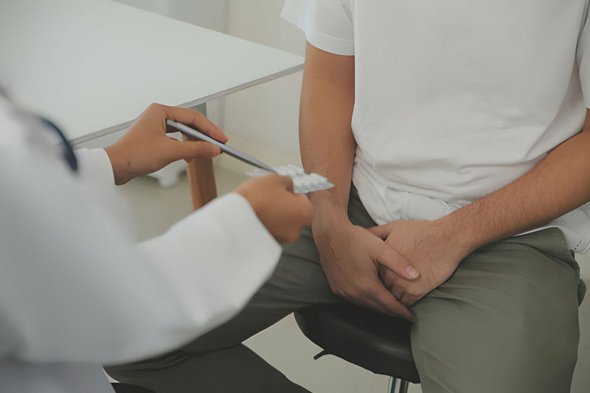
Undescended testicles may occur in 3-4 of every 100 newborns, and it is more common in premature babies. Testicular surgery is a successfully applied method for the treatment of this disease.
In half of the babies with undescended testicles in the neonatal period, the testicles descend into the scrotum within the first three months. Babies 6-12. If they still have undescended testicles when they reach the first month of life, the testicles are no longer expected to descend into the scrotum. 1 or 2 in 100 children with undescended testicles need treatment.
It is important to distinguish between undescended testicles and retractile testicles. In a 6-month-old male baby, the testicles may temporarily shift upwards as a reflex that develops due to cold or fear. Normally, the testicles in the scrotum do not require treatment. In the presence of truly undescended testicles, treatment is required. This condition can be easily distinguished during examination by a urologist.
In the presence of non-palpable or undescended testicles in newborns and if sexual development disorders such as hypospadias are detected, urgent endocrinological and genetic examination must be performed.
In order for the testicles to produce sperm, their temperature must be 2-3 degrees lower than body temperature. The scrotum is at a lower temperature than body temperature and is the ideal place for the testicles. Therefore, testicles that do not descend into the scrotum may have a defect in sperm production. As the testicles remain in a warm environment, the chance of normal development of sperm decreases.
In cases where bilateral testicles are affected, infertility may be an expected result. In cases with undescended testicles, the incidence of testicular cancer and testicular torsion, as well as infertility, is higher than in the normal population. This situation reveals how important it is to recognize and treat undescended testicles early.

What is Undescended Testis (Cryptorchidia)?
Normal testes are formed in the male fetus in the womb during the early period of development. They form in the lower abdomen of the fetus, and as the pregnancy progresses, they move downwards and descend into the scrotum. In detailed ultrasound performed during pregnancy, the descent of the testicle cannot be observed until the 28th week, only its movement from the abdomen to the internal inguinal ring can be seen.
28-40 due to the effect of secreted hormones. Between weeks of pregnancy, the descent of the testicles into the scrotum is completed. The testicles are attached to the lower part of the scrotum through stretchable tissues.
True undescended testicle is the condition in which the testicle is on the path required for it to descend to the scrotum during development in the womb, but cannot reach the scrotum. Undescended testicles are classified according to the location of the testicles and their palpability during examination.
Undescended Testicular Cryptorchid Types
Undescended testicles are primarily evaluated in two groups: palpable (palpable during examination) and non-palpable. 80% of undescended testicles can be palpated. Treatment and follow-up for undescended testicles vary depending on the location and condition of the testicles.
A. Palpable (palpable on examination) testicles
True Undescended Testicle
Ectopic Testis
B. Non-palpable (not palpable on examination) testicles
Testes located in the abdomen: They constitute 50-60% of the non-palpable testicles. They are often located close to the internal inguinal ring, but can also be around the kidney, around the anterior abdominal wall and behind the urinary bladder.
Inguinal located testicles
ectopic testicle
Absence of testicles (testicular agenesis and vanishing testicle syndrome)
Who gets undescended testicles?
Cryptorchidism, or undescended testicle, is the most common congenital anomaly in newborn male babies. The incidence of undescended testicles varies depending on the week of pregnancy in which the babies are born.
While the incidence of undescended testicles is 1-4.6% in term babies, this rate increases to 1.1-45% in premature babies.
In term babies, the incidence of undescended testicles is 1% in male babies who are 1 year old, even after the testicle spontaneously descends into the scrotum in the first month after birth. In 30% of cases, both testicles may be affected. It is not an expected situation for the undescended testicle to spontaneously descend into the scrotum after the age of 1. Common conditions for undescended testicles include:
prematurity
low birth weight
Being underweight for gestational age
Maternal use of estrogen in the first trimester of pregnancy
Family history of undescended testicles

How Is Undescended Testicle Diagnosed?
For the diagnosis of undescended testicle, taking a detailed history and physical examination of the patient is very important. The child’s detailed birth history (birth weight and week), whether the mother received hormonal treatment before and during pregnancy, and whether there is a history of undescended testicles or another sexual disorder in the family should be questioned. For detailed information, you can visit our sexual health center page.
During the physical examination of the patient, an attempt is made to detect the testicle by manual examination from the groin to the scrotum. It is examined whether there is a scrotal disorder.
The patient’s other existing testicle is examined, and if it is hypertrophic, this suggests testicle absence or atrophy. Imaging methods such as ultrasound or MRI cannot give a definitive result as to whether the testicle is present or not.
The correct diagnosis rate of undescended testicle with imaging methods is 44%. Physical examination performed by a urologist is more valuable than imaging methods for diagnosis.